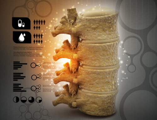

The spine consists of a complex system of bones and ligaments that provide stability and support for the entire body. This complex system contains a spinal column that protects the spinal cord and nerves. The spinal cord and nerves transmit signals from the brain to the rest of the body. If the bones or tissues surrounding the spinal cord are damaged or narrowed, it can affect the space around the spinal cord and disrupt basic movements like walking, balance, and sensation.
Spinal stenosis is a condition that causes the spinal column to narrow and compresses the spinal cord. This compression typically develops gradually, with a slow progression of symptoms. If the space around the spinal cord eventually becomes too narrow, it can affect the transmission of signals from the brain through the spinal cord.
While aging is the most common cause, conditions like osteoarthritis, disc herniations, ligament thickness and rheumatoid arthritis can contribute to the pressure on your spinal cord.
What are the Symptoms of Spinal Stenosis
There are common symptoms related to spinal stenosis, each dependent on the location of the stenosis. Spinal stenosis symptoms typically have a gradual onset, starting slowly and getting worse over time. Spinal stenosis may appear on an MRI or a CT scan without the patient having any symptoms at all.
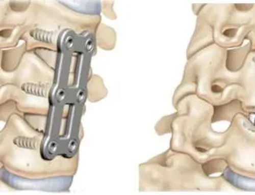
Cervical Spine
- Numbness or tingling in the hand, arm, foot, or leg
- Difficulty walking or with balance
- Neck pain
- Bowel or bladder conditions such as urinary urgency or incontinence
Lumbar Spine
- Weakness in the foot or leg
- Pain or cramping in one or both legs after long periods of standing or walking
- Back pain
- Numbness or tingling in the leg or foot
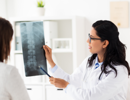
How Is It Diagnosed?
Your doctor will diagnose this condition by reviewing your medical history and perform a physical examination. They will also want to discuss any related symptoms you experience. After a full review, your doctor may order several imaging tests to pinpoint the cause of your symptoms.
Imaging tests may include:
X-Rays
X-rays can determine if there was a bone change that could affect the spinal column, such as bone spurs or instability.
Magnetic Resonance Imaging (MRI)
MRIs utilize a powerful magnet and cross-sectional radio waves to produce images of your spine. MRIs detect issues with ligaments and discs in addition to evaluating the pressure on the nerves. MRIs are helpful in determining the location and severity of spinal stenosis. Your doctor will be able to evaluate your symptoms and the MRI together to determine the best treatment options.
CT Scan
If you are unable to have an MRI, a CT scan may be used instead. This type of imaging test combines X-Ray images that are taken at many different angles to create a detailed, cross-sectional image of your body. A CT/Myelogram may be indicated to demonstrate the extent of spinal stenosis and the exact location of compression on the spinal cord or nerves.
The team at First State Spine can answer any questions you may have about spinal stenosis and is trained in the diagnosis and treatment of this condition. Contact them today!
Sources: